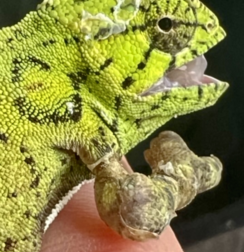
Skin Deep and Scale Tight: Shedding Disorders in Chameleons and the Anatomy Behind the Crisis

Reptilian skin is a highly specialized organ system, serving as both a protective barrier and a dynamic interface with the environment. In chameleons, the skin is composed of two principal layers. The outer layer, the epidermis, includes the stratum germinativum, which is mitotically active and responsible for generating new cells, and the stratum corneum, a keratinized, anucleate layer that is periodically shed. Beneath the epidermis lies the dermis, which contains connective tissue, chromatophores, vascular and neural networks, and exocrine glands. Chameleons possess an advanced dermal architecture that supports rapid and complex color modulation through iridophores and melanophores, enabling visual signaling, camouflage, and thermoregulation (Tolley & Herrel 2013; Teyssier & al. 2015).
Despite their often miniature size, chameleons possess a complete suite of vertebrate organ systems. Even the smallest species, such as Brookesia minima, measuring just over 3 cm in total length, contain fully functional hearts, livers, kidneys, lungs, endocrine glands, and central nervous systems. These organs are not simplified but scaled with astonishing precision. The presence of microscopic glands and tightly regulated metabolic and hormonal systems in such minute bodies is a testament to the anatomical and physiological sophistication of these animals (Nečas 1999).
Shedding, or ecdysis (from Greek ἐκδύειν, "to strip off"), is the process by which reptiles replace the outer layer of their epidermis. In chameleons, shedding is primarily a function of growth and secondarily of skin renewal. Juveniles may shed up to twice weekly due to rapid somatic expansion, while adults typically shed once or twice per year depending on species, environmental conditions, and individual health status (Lillywhite 2006).
Chameleons are classified as dry shedders. The shedding process begins with lymphatic fluid infiltrating the space between the old and new epidermis, initiating separation. However, this fluid evaporates quickly, leaving a thin layer of air between the two skins. Under optimal conditions—moderate humidity, stable temperatures, and access to basking—the skin may be shed in large, contiguous sheets. Juveniles and small-bodied species often shed in one piece. Larger species may do so as well if conditions are ideal. When environmental parameters are suboptimal—excessive moisture, low temperatures, or prolonged rainfall—capillary action may draw water into the inter-epidermal space. This causes the layers to adhere, resulting in dysecdysis (Mader 2005).
The most serious complication arises when retained epidermal rings form around extremities, particularly the elbows, metacarps, metatarsals, forearms, tibias, and thighs. In adults, these constrictions may remain subclinical for extended periods. In juveniles, however, they pose a significant threat. Because retained skin cannot be shed again, it forms a tight band that impedes vascular and lymphatic circulation. As the limb grows, the constriction intensifies, leading to edema, ischemia, necrosis, and potentially sepsis. If untreated, this can result in permanent tissue damage, autoamputation, or death (Divers & Mader 2005).
In wild populations, shedding complications are rarely documented. Affected individuals often perish unnoticed, their remains lost to decomposition or predation. In captivity, however, intervention is both possible and necessary. Regular monitoring is essential. Retained skin should be moistened with isotonic antiseptic solution, such as diluted chlorhexidine or sterile saline, and gently removed with sterile forceps. If the site exhibits signs of infection or tissue compromise, topical antibiotics such as silver sulfadiazine should be applied, and systemic therapy considered under veterinary supervision (Wilkinson 2008).
This condition underscores the importance of precise husbandry, environmental control, and routine clinical inspection in captive chameleon management. It also highlights the evolutionary elegance of these animals, whose anatomical and physiological complexity rivals that of any vertebrate, regardless of size.
References
Alibardi, L. 2003. Adaptation to the land: the skin of reptiles in comparison to that of amphibians and mammals. Journal of Experimental Zoology Part B: Molecular and Developmental Evolution 298: 12–41.
Divers, S.J. & Mader, D.R. 2005. Reptile dermatology. In: Mader, D.R. (ed.) Reptile Medicine and Surgery. 2nd ed. St. Louis: Saunders. 1242pp.
Lillywhite, H.B. 2006. Physiology and functional morphology of reptilian skin. In: Gans, C.; Gaunt, A.S. & Adler, K. (eds.) Biology of the Reptilia. Vol. 20. Ithaca: Society for the Study of Amphibians and Reptiles. 687pp.
Mader, D.R. 2005. Reptile Medicine and Surgery. 2nd ed. St. Louis: Saunders. 1242pp.
Maderson, P.F.A. 1985. Some developmental problems of the reptilian integument. In: Gans, C. & Billett, F. (eds.) Biology of the Reptilia. Vol. 14. New York: Wiley. 624pp.
Nečas, P. 1999. Chameleons: Nature's Hidden Jewels. Frankfurt am Main: Edition Chimaira. 348pp.
Teyssier, J.; Saenko, S.V.; van der Marel, D. & Milinkovitch, M.C. 2015. Photonic crystals cause active colour change in chameleons. Nature Communications 6: 6368.
Tolley, K.A. & Herrel, A. 2013. The Biology of Chameleons. Berkeley: University of California Press. 288pp.
Wilkinson, R. 2008. Clinical pathology. In: Doneley, B.; Monks, D.; Johnson, R. & Carmel, B. (eds.) Reptile Medicine and Surgery in Clinical Practice